Blog Details
Table of Contents
- What AI Skin Diagnostics Actually Means
- How AI Skin Diagnostic Tools Capture and Process Images
- Pattern Recognition: How AI Identifies Skin Characteristics
- AI Skin Analysis Accuracy: What the Data Shows
- AI Skin Cancer Detection and Clinical Validation
- Using AI Skin Analysis for Personalised Treatment Planning
- Before, During and After: What Practitioners and Patients Should Know
- Frequently Asked Questions
Last Updated: October 12, 2026
What AI Skin Diagnostics Actually Means
AI skin diagnostics uses artificial intelligence and machine learning to analyse skin images and identify concerns, conditions, or changes warranting clinical attention. Computer vision detects patterns in colour, texture, shape, and symmetry the human eye might miss.
The key distinction: AI skin diagnostics supports clinical decision-making; it does not replace a dermatologist’s judgment.
At DermTech Devices, we understand modern aesthetic practices need reliable, objective data to justify treatment decisions and track progress. AI skin analysis turns subjective assessment into measurable baseline data, making results easier to demonstrate.
The process works in stages: image capture, standardisation, analysis through trained neural networks, pattern recognition, and a report highlighting areas of concern.
The most common mistake is assuming AI skin diagnostics is a diagnostic tool in the medical sense. It is not. It is an analysis tool that flags patterns and supports your clinical reasoning. Always correlate AI findings with your own clinical assessment and patient history.
How AI Skin Diagnostic Tools Capture and Process Images
Capturing a usable skin image is more technical than pointing a camera at someone’s face. Standardisation matters enormously because the AI model was trained on images captured under consistent conditions.
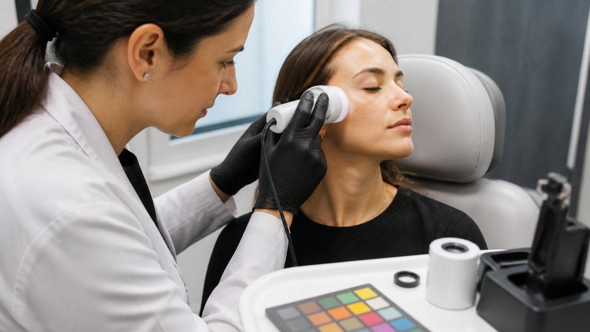
Most professional systems use handheld imaging devices or standardised smartphone capture with calibration markers. The device captures multiple wavelengths, visible, polarised, sometimes UV, to reveal different skin layers.
Image standardisation is critical. The system needs:
- Consistent distance from skin (typically 5-10 cm)
- Standardised lighting (usually diffuse, shadow-free)
- Colour reference cards in frame for calibration
- Multiple angles or zones of the same area
- Patient positioning that minimises movement
Software then corrects lighting variation, standardises colour space, and removes artifacts before the image enters the AI pipeline.
Machine learning and deep learning models
The AI backbone uses deep learning, specifically convolutional neural networks (CNNs), trained on large datasets of skin images with known clinical outcomes, learning patterns linked to hyperpigmentation, texture irregularities, lesion morphology, and vascular patterns.
Training data is crucial. A model trained only on lighter skin tones performs poorly on deeper tones. Responsible systems use diverse datasets tested across skin tone ranges.
The deep learning model does not “think” like a dermatologist. It finds statistical patterns in pixel data and, when those correlate with a clinical outcome, flags them with confidence scores.
Machine learning models are only as good as their training data. A model trained on 10,000 images of fair skin will not reliably analyse darker skin tones. Always check whether your AI system has been validated across the patient populations you treat.
Pattern Recognition: How AI Identifies Skin Characteristics
Once the image is standardised, the AI looks for patterns. It does not see a “face”, it sees pixel values, colour distributions, and texture gradients, and has learned which combinations correlate with specific conditions.
Analysing colour, texture, shape and symmetry
Colour analysis detects melanin distribution, erythema (redness), and vascular patterns.
Texture analysis examines surface roughness, pore size, and fine lines by mapping micro-topography from local brightness variations.
Shape and symmetry assessment looks at lesion borders, size, and asymmetry. Melanoma screening systems focus on border irregularity and asymmetry, clinical red flags, measuring them objectively rather than relying on variable visual judgment.
The system also detects subcellular characteristics when imaging technology allows.
Detecting lesions and cellular patterns
Lesion detection works through segmentation: the algorithm identifies where a lesion begins and ends, separates it from surrounding skin, and quantifies size, border definition, colour variation, and asymmetry for risk stratification.
For skin cancer screening, the system flags lesions matching patterns associated with melanoma or other malignancies.
Cellular pattern recognition looks at deeper skin layers when imaging permits. Atypical keratinocytes, abnormal melanocytes, and inflammatory infiltrates leave signatures the AI learns to flag as potential indicators of dysplasia or malignancy.
AI Skin Analysis Accuracy: What the Data Shows
Accuracy in AI skin diagnostics is measured by sensitivity (true positives caught) and specificity (true negatives correctly identified). High sensitivity catches most real cases but may flag false alarms; high specificity means fewer false alarms but risks missing real cases.
Sensitivity, specificity and false positives
No AI system is perfect. A false positive flags something concerning when it is benign; a false negative misses something that is actually concerning.
For skin cancer detection, missing a melanoma is worse than a false alarm leading to biopsy. Most published studies show well-trained AI systems achieve 85-95% sensitivity on curated datasets, though real-world performance can be lower because clinical images are messier (pubmed.ncbi.nlm.nih.gov).
Specificity ranges widely: high against obviously benign nevi, but lower against atypical nevi because the distinction is harder.
The practical implication: use AI as a screening and support tool, not a diagnostic tool. Take high-risk flags seriously; if the system rates something low-risk, still examine the lesion yourself.
Performance across skin tones
This is where AI skin diagnostics reveals its limitations.
Recent systems are being retrained with more diverse datasets, and responsible vendors now publish performance breakdowns by skin tone. Ask for this data when evaluating a system; if unavailable, be cautious.
The concern is not theoretical: melanoma is often diagnosed later in patients with darker skin tones, partly because clinical tools (including AI) have been optimised for lighter skin. A poorly performing system could perpetuate that disparity.
Never assume an AI skin diagnostic system performs equally across all skin tones. Ask your vendor for validation data broken down by Fitzpatrick skin type or similar classification. If they cannot provide it, the system has not been adequately tested on diverse populations.
::: (Source: ethical considerations for AI in healthcare)
AI Skin Cancer Detection and Clinical Validation
Skin cancer detection is the most high-stakes application of AI skin diagnostics. The goal is to identify lesions that warrant biopsy or referral before they progress.
Melanoma and common skin cancers
Melanoma is the most aggressive skin cancer, and early detection dramatically improves prognosis. AI systems trained to detect it look for the ABCDE criteria, asymmetry, border irregularity, colour variation, diameter over 6 mm, and evolving appearance, plus other learned patterns (Melanoma Treatment (PDQ®)).
Basal cell carcinoma (BCC) and squamous cell carcinoma (SCC) are more common but less aggressive. They present with different visual patterns: BCC often shows a pearly appearance and telangiectasia; SCC may appear scaly or ulcerated.
The system quantifies these features objectively. Instead of one practitioner saying “this looks suspicious” and another “it looks fine,” the AI produces a risk score based on measurable features, reducing observer variation and creating a baseline for tracking changes.
Supporting clinical diagnosis, not replacing it
This cannot be overstated: AI skin diagnostics supports your clinical judgment. It does not replace it.
The AI system sees patterns.
A responsible workflow integrates AI findings with clinical assessment. If the AI flags a lesion high-risk but your judgment and dermoscopy suggest it is benign, trust your judgment; if the AI rates it low-risk but you have concerns, investigate further.
Professional bodies and regulatory frameworks emphasise this: AI enhances clinical decision-making, it does not replace the clinician’s responsibility.
Using AI Skin Analysis for Personalised Treatment Planning
Beyond cancer screening, AI skin analysis generates objective baseline data that transforms treatment planning and outcome tracking.
Objective data for treatment decisions
Before treatment, capture baseline images with the AI system, which quantifies skin texture, pigmentation, vascularity, and other parameters to create a measurable starting point.
When deciding on a protocol, laser resurfacing, IPL, chemical peels, or other modalities, you can reference objective data. “Your baseline texture score is 7.2” is more concrete than “your skin looks rough,” and patients feel more confident in treatment decisions.
Objective baseline data also protects your practice. If a patient later disputes whether they improved, you have quantitative before-and-after data. This reduces disputes and supports your clinical record.
Tracking progress and adjusting protocols
Capture follow-up images at regular intervals, typically 4-8 weeks depending on treatment.
This data guides protocol adjustment: a good response after one laser session may mean continuing the same protocol, while minimal response may mean adjusting power, frequency, or switching modalities. Objective data makes these decisions more evidence-based.
For practices using DermTech Devices systems, this objective tracking becomes part of your standard workflow. Patients see their own progress data, building confidence and retention, while your team documents outcomes systematically to strengthen case studies and marketing.
Before, During and After: What Practitioners and Patients Should Know
Successful AI skin analysis depends on proper preparation, correct technique during capture, and informed interpretation of results.
Preparing for a skin scan
Position the patient comfortably with adequate lighting, most professional systems include lighting modules, so you do not rely on room lighting. Explain that the system captures multiple images from different angles in just a few minutes.
Ensure the imaging device is calibrated. Most systems require a daily or weekly check using a reference standard; skipping it introduces error into your baseline data.
Understanding results and next steps
The AI system produces a report highlighting areas of concern, assigning risk scores, and comparing current findings to baseline where prior images exist.
Review the report with the patient. Explain what the scores mean. “Your pigmentation score improved by 12% since your last visit” is actionable feedback.
If the system flags a lesion as high-risk for skin cancer, explain that it warrants closer clinical assessment, dermoscopy, biopsy, or specialist referral.
For cosmetic concerns, use the data to set realistic expectations: “Your baseline texture score is 6.8.
Privacy and consent in image handling
Skin images are sensitive personal data. Ensure your practice has clear consent processes and secure storage.
Patients should understand:
- What images will be captured and why
- How images will be stored and for how long
- Who can access images (your staff, the AI vendor, anyone else?)
- Whether images might be used for training or research
- Their rights to request deletion
Document consent in your patient records. Store images securely, encrypted, access-controlled, backed up. If you use cloud-based AI systems, understand the vendor’s data handling practices. Do they retain images?
Regulatory frameworks like the Privacy Act 1988 (Cth) in Australia govern how health information must be handled. Comply with these requirements. Your practice’s reputation depends on protecting patient data.
Objective skin analysis transforms how aesthetic and dermatology practices make treatment decisions and track progress.
At DermTech Devices, we build AI skin analysis systems designed for clinical reliability and ease of integration into your workflow. We partner with you to implement objective data capture as standard practice, supporting better treatment outcomes and stronger practice growth. Partner with us to bring objective, AI-powered skin analysis into your clinic.
Frequently Asked Questions
How does AI skin diagnostics work?
AI skin diagnostics analyse digital images of skin using machine learning algorithms trained on thousands of images. The system identifies patterns in colour, texture, shape and symmetry to assess skin conditions and concerns. Computer vision technology processes the image data and compares it against trained models to generate a risk assessment or diagnostic score. The result supports clinical decision-making but does not replace professional diagnosis by a dermatologist or qualified practitioner.
How accurate is AI skin analysis?
AI skin analysis accuracy varies by condition and algorithm. For melanoma detection, validated systems report sensitivity (true positive rate) between 85-95% and specificity (true negative rate) between 75-90%, though performance depends on training data diversity and real-world testing. Accuracy is higher when images are well-lit, standardised and captured on diverse skin tones. No AI system is 100% accurate; false positives and false negatives occur, which is why clinical validation and physician review remain essential.
Can AI detect skin cancer?
AI can identify features consistent with melanoma and other common skin cancers, including irregular borders, colour variation and asymmetry. However, AI functions as a screening and decision-support tool, not a diagnostic device. A qualified dermatologist or trained practitioner must review results and perform clinical examination. AI excels at flagging lesions that warrant closer attention, reducing missed cases and supporting faster referral when needed.
How should practitioners use AI skin analysis results in treatment planning?
Use AI analysis data as objective baseline measurements to inform treatment selection, set realistic expectations and track progress over time. For aesthetic concerns, the analysis identifies priority areas (pigmentation, texture, hydration, fine lines) to guide protocol choice and intensity. Document results before and after treatment to demonstrate efficacy to patients and refine your protocols. Combine AI data with clinical assessment and patient goals to create personalised treatment plans rather than relying on AI results alone.