
Blog Details
Last Updated: November 20, 2026
Quick Summary
Skin analysis technology for acne assessment uses facial images and software to map lesions, measure redness and texture, and track change over time. It makes grading more consistent, but automated results should supplement, not replace, a clinician’s examination and diagnosis.
- Lighting, camera distance, makeup and facial hair can all distort an acne scan.
- Grade lesions by type, since comedones, papules, pustules and nodules differ clinically.
- Read the severity band as the headline and the score as a trend line.
- Repeat scans under identical conditions to make progress comparisons meaningful.
- See a dermatologist if acne is severe, painful, scarring, spreading or not improving.
Table of Contents
- What Skin Analysis Technology Does and How It Works
- How Acne Is Graded and What Scores Actually Mean
- AI Skin Analysis for Acne: Detection and Lesion Mapping
- AI Acne Grading: Standardising Assessment Across Settings
- Skin Analysis Device Accuracy, Skin Tones and Scan Distortion
- Tracking Acne Treatment Progress With Repeat Scans
- Booking Your Acne Assessment and When to See a Dermatologist
- Frequently Asked Questions
Acne is one of the most common reasons people visit a skin clinic, and assessing it accurately has always depended on how the practitioner looks at the skin. Skin analysis technology for acne assessment aims to make that judgement more consistent, using facial images and software to map lesions and track change over time. At DermTech Devices, we build skin analysis systems for dermatology and aesthetic practices, so we have a direct interest in how these tools are used and where their limits lie. This guide explains what the technology does, how acne severity is graded, and where automated results should give way to professional care. (Source: Therapeutic Goods Administration)
What Skin Analysis Technology Does and How It Works
Skin analysis technology captures images of the face under controlled conditions and uses software to measure visible features such as redness, texture, pigmentation and lesions. See how practitioners apply these tools in clinical workflows. You can compare the range of available skin analysis equipment to see what each category offers. Most systems compare these measurements against reference data and present the results as scores or annotated images. The aim is an objective record that can be compared from one visit to the next.
Some consumer tools work from a simple selfie and are designed for skincare product recommendation, which is a different job from clinical assessment.
How Acne Is Graded and What Scores Actually Mean
A severity score is only useful if you know what went into it. Acne grading generally follows one of three routes: a global severity grade assigned by a clinician, a manual count of individual lesions, or multimodal digital imaging that combines several image types. Each produces a different kind of number, and they are not interchangeable.
The lesion types a score should reflect
Any credible assessment starts by separating lesions into types, because they behave differently and respond to different treatments:
- Comedones, open and closed plugs (blackheads and whiteheads) that are non-inflammatory.
- Papules and pustules, small inflamed bumps, with pustules containing visible pus.
- Nodules and cysts, deeper, larger, often painful lesions that carry a higher scarring risk.
A count that lumps all of these together can make two very different faces look identical on paper. A patient with 20 comedones and a patient with 20 nodules are not at the same point, even if the total is the same.
Distribution and extent matter as much as the count
Where acne sits changes its clinical meaning. Jawline and chin patterns are common in hormonal presentations, while widespread involvement across cheeks, forehead and trunk suggests a broader process. Good grading therefore records the regions involved and the proportion of skin affected, not just a total. When you review a report, check whether it breaks results down by facial zone, a single whole-face number hides the pattern that guides treatment.
Reading the number in context
Most systems present results as a severity band (for example, clear, mild, moderate or severe) alongside a numeric score. Treat the band as the headline and the number as a trend line. A move from 42 to 38 means little on its own; a move from moderate to mild, sustained across two or three scans under the same conditions, is the kind of change worth discussing with your clinician.
Watch out: a lower score is not automatically an improvement if the mix of lesion types has shifted toward deeper nodules, so always read the score alongside the lesion breakdown.
Automated estimates versus clinician assessment
Automated grading estimates what a camera can see. A clinician assesses what the camera cannot: how deep a lesion feels, whether it is tender, how long it has been present, scarring history, and the person’s response to previous treatments. The most reliable picture combines both, the scan supplies consistency and a visual record, and the clinician supplies diagnosis and judgement. Ask your clinic which grading method its system uses and whether that method matches the scale used to plan your treatment.
AI Skin Analysis for Acne: Detection and Lesion Mapping
AI-based systems try to detect and map acne lesions across the face from a photograph. Research has explored AI-powered facial analysis for detecting acne types and oiliness, and results should be checked in the full published papers before you rely on them. For a deeper clinical explanation of how AI diagnostics detect and map lesions, read how AI skin diagnostics work.
Mapping lesions by region is where these tools can add the most value, because it shows whether acne is concentrated on the jawline, the cheeks or the forehead. That distribution is harder to record consistently by eye across repeat visits.
AI Acne Grading: Standardising Assessment Across Settings
One reason for interest in AI grading is consistency. Some commentary suggests that AI-based grading could help standardise assessment across settings. If practitioners and researchers apply the same method, results become easier to compare.
Standardisation is still a goal rather than a settled fact. Two clinics using different tools may still get different results, so it helps to confirm which grading method a system uses and whether it matches the scale your practice or study relies on.
Skin Analysis Device Accuracy, Skin Tones and Scan Distortion
Accuracy is not a fixed property of a device, it is the product of the hardware, the software and the conditions of each scan. Learn how objective skin analysis is applied in aesthetic clinics. Two clinics can run the same system and get different results if their capture conditions differ. Understanding what moves the result is the fastest way to make scans comparable.
The factors that distort an acne scan
Most misleading results trace back to a short list of variables:
- Lighting, uneven light, shadows and mixed colour temperature change how redness and texture are detected. Consistent, diffuse lighting is the single biggest lever.
- Camera and distance, different cameras, lenses and focal lengths render pores and lesions differently. Keep the same device and the same distance from the face.
- Makeup and skincare, foundation, tinted sunscreen and occlusive products can mask lesions or alter surface shine. Scan clean skin where possible.
- Facial hair, stubble and beards obscure the jawline and chin, which are common acne sites, so note hair status at each scan.
- Oil and shine, recent exercise, heat or heavy moisturiser can create specular highlights that software misreads as texture.
- Angle and expression, a tilted head or a different expression shifts the regions captured. Standardise pose and expression.
Watch out: scanning straight after cleansing can make skin look temporarily calmer, so allow a consistent settling period before every scan.
A repeatable preparation routine
A practical workflow is to scan at the same time of day, with clean dry skin, no makeup, the same device and distance, and the same head position. Record the date, the conditions and any changes to products or medication alongside the images. That record is what turns a series of scans into a usable trend rather than a set of unrelated pictures.
Performance across skin tones
Colour and contrast affect how lesions, redness and post-inflammatory marks appear in an image, and many systems were trained on datasets that were not evenly balanced across skin tones. Performance can therefore vary by phototype, particularly for detecting redness-based features on deeper skin. Public validation data on this point is uneven between products, so ask suppliers for testing that reports results by skin tone rather than an overall accuracy figure. A single headline accuracy number tells you little about how the system will perform on the patients in front of you.
Privacy and facial-image data
Selfie-based and clinic-based tools both process sensitive facial images, which may be treated as personal information under Australian privacy law. Before a scan, ask where the images are stored, how long they are retained, whether they are used to train models, and how consent is recorded. A clinic that cannot answer these questions clearly is a reason to pause.
Questions worth asking a supplier
Ask which grading method the system uses, whether validation results are reported by skin tone, what capture conditions the accuracy claims assume, and how images and derived data are stored and deleted. Documented answers to those four questions separate a clinical tool from a marketing demo.
The main variables that can distort an acne scan, and what to do about each, are summarised below.
| Factor | Why it distorts the result | Fix |
|---|---|---|
| Lighting | Shadows and mixed colour temperature change detected redness and texture | Use consistent, diffuse lighting |
| Camera and distance | Lenses render pores and lesions differently | Keep the same device and distance |
| Makeup and skincare | Foundation and occlusive products can mask lesions or add shine | Scan clean skin where possible |
| Facial hair | Stubble and beards obscure the jawline and chin | Note hair status at each scan |
| Oil and shine | Specular highlights read as texture | Allow a consistent settling period |
| Angle and expression | Shifts the regions captured | Standardise pose and expression |
Tracking Acne Treatment Progress With Repeat Scans
Repeat scans are most useful when the conditions are kept identical each time. Use the same lighting, the same camera setup, the same time of day and a similar expression and pose. Record the lesion counts and regions alongside the scores, so a change can be read in context rather than as a single number.
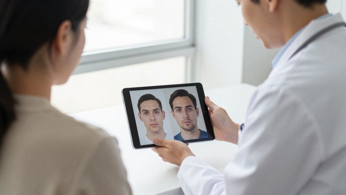
Progress tracking helps show whether a treatment plan is working, but a change in score is a prompt for discussion with the treating clinician, not a verdict on its own.
Booking Your Acne Assessment and When to See a Dermatologist
If you are considering an acne assessment, a consultation with a qualified dermatologist or skin professional is the right first step. To compare the skin analysis systems offered by clinics, review the options before you book. Automated analysis can support that conversation, but it does not replace a clinical examination, and it cannot diagnose the cause of your acne or tell you which treatment is appropriate for you.
See a dermatologist sooner rather than later if acne is severe, painful, scarring, spreading quickly, or not improving with over-the-counter care. Ask your clinic how its assessment is carried out, which grading method it uses, and whether repeat scans will be compared under consistent conditions. For more on how DermTech Devices supports practices with skin analysis systems, get in touch with our team to talk through your options.
Frequently Asked Questions
How does skin analysis technology assess acne?
Most systems capture standardised facial photographs and use image analysis to identify and count visible lesions, then estimate where on the face they cluster. Research on acne grading describes three broad approaches: global severity grading, lesion counting, and multimodal digital imaging. A good system should record lesion type, number and distribution, not only a cosmetic skin score. Results support, rather than replace, a clinical assessment by a qualified practitioner.
Can AI skin analysis identify different types of acne?
Some AI tools aim to distinguish acne types and oiliness from facial images, and published research has explored this for personalised product suggestions. Clinically, acne is described by primary lesion types such as comedones, papules, pustules and nodules, which differ in severity. Whether a given tool separates these reliably depends on its validation, so ask the supplier for evidence on the specific lesion categories it reports.
How accurate are skin analysis devices for acne assessment?
Accuracy depends on the device, image quality, lighting and the population it was validated on. Many consumer and commercial tools have not been independently validated for acne grading, so their output should be treated as a guide. Accuracy claims should be checked against peer-reviewed validation data, including performance across different skin tones. Clinical judgement remains the reference standard for diagnosis and treatment decisions.
Can skin analysis technology track acne treatment progress?
Repeat scans taken under the same conditions can show changes in lesion counts and distribution over time. To make comparisons meaningful, keep camera distance, lighting, skin preparation and timing consistent between sessions. Progress tracking is most useful when reviewed alongside a dermatologist or treating clinician, who can judge whether changes reflect true improvement.